

Microscopic Root Canal Treatment in Bangalore
Unparalleled precision in endodontic care. We use advanced dental operating microscopes to find hidden canals and ensure maximum clinical success.
The Pinnacle of Precision: Microscopic Endodontics
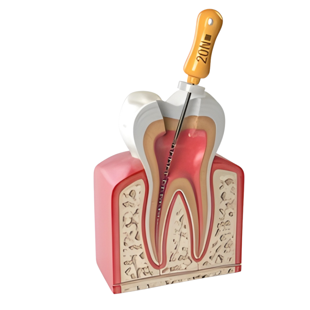
Root canal treatment requires working inside an incredibly small, dark, and complex space—the inside of your tooth. The root canals themselves are often narrower than a human hair and can feature complex curves, branches, and hidden anatomical structures. Performing this delicate work using only the naked eye or basic magnifying loupes leaves a margin for error. At Arkas Dental Studio in KR Puram, Bangalore, we elevate our endodontic care by offering Microscopic Root Canal Treatment, utilizing state-of-the-art Dental Operating Microscopes (DOM).
A dental operating microscope provides brilliant, coaxial illumination (light shining directly down the line of sight) and extreme magnification (up to 25x). This allows our specialists to see deep inside the root canal system with unprecedented clarity. The difference between traditional RCT and microscopic RCT is profound: it is the difference between feeling your way in the dark and working in a brightly lit room.
Why Magnification Matters in Root Canals
The use of a microscope significantly increases the long-term success and predictability of root canal therapy by allowing the dentist to:
- Locate Hidden Canals: Many teeth (especially molars) have extra, hidden canals that are invisible to the naked eye. If missed, the infection remains, causing the root canal to fail. The microscope ensures every canal is found and cleaned.
- Identify Micro-Fractures: Tiny cracks in the tooth structure can cause severe pain and doom a tooth to extraction if not identified. Magnification allows us to spot these micro-fractures early.
- Conserve Tooth Structure: Because we can see exactly where we are drilling, we remove far less healthy tooth structure to access the canals, leaving the tooth much stronger and less prone to fracture later.
- Navigate Complex Anatomy: Highly calcified (blocked) or severely curved canals can be negotiated and cleaned safely without accidentally perforating the root.
When is Microscopic RCT Recommended?
While we believe the enhanced vision of a microscope benefits every endodontic procedure, it becomes absolutely essential in several complex clinical scenarios:
Molar Treatments
Back teeth (molars) inherently have complex anatomies, often possessing three, four, or even five canals, some of which are exceptionally fine. The microscope is vital for successfully treating these teeth.
Root Canal Retreatment
If a previous root canal has failed and become re-infected, treating it again (retreatment) is much more difficult. The old filling material must be removed, and the cause of the failure (such as a missed canal or separated instrument) must be addressed. The extreme magnification of a microscope is mandatory for a successful retreatment.
Managing Complications
In cases where a previous dentist may have accidentally broken a small file inside the canal, or created a perforation in the root wall, microscopic endodontics allows our specialists to safely bypass or remove the obstruction and repair the perforation, saving a tooth that would otherwise need extraction.
Advanced Precision Care
Ensure the highest success rate for your root canal with our advanced microscopic technology.
Why Arkas Dental Studio?
Experienced Professionals
Predictable, comfortable outcomes.
Advanced Technology
Modern clinical tools.
Strict Sterilization
Patient safety comes first.